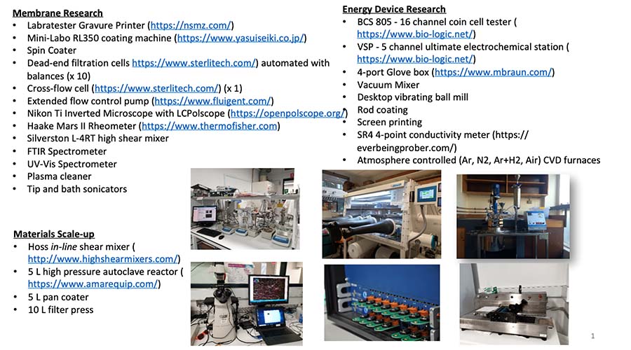
Facilities
MATERIALS SYNTHESIS & PROCESSING EQUIPMENT
-
- Fully serviced fume cupboards
- Chemical vapour deposition reactors up to 1400°C
- Plasma etcher
- Variable speed spin coater
- Automated gravure coater
- Bath and tip-ultrasonicator
- Centrifuge
- Acid-resistant chemical reactors (5 L) for upscaling Graphene synthesis
- Computer controlled pulsed UV light source
CHARACTERIZATION EQUIPMENTS FOR ENERGY, CHEMICAL SEPARATIONS AND MICRO-/NANO-FLUIDIC APPLICATIONS
- Multichannel BioLogic Potentiostat (currently 4 active channels installed)
- Solar simulator
- Newport vibration isolation table with Genelyte Mini probe station from EmCal
- Ocean Optics UV-vis spectrometer
- Agilent B2902 A and Keithley 6430 sub femto-Amp SMU
- High pressure (20 bar) dead-end membrane filtration
- Packed bed adsorption columns
- Nikon Eclipse Ti inverted microscope
- C2V Portable Gas Chromatograph
- Scanning Ion Conductance Microscope (IC Nano S2 system): Scanning Ion Conductance Microscopy (SICM) acquires topographic images of surfaces in electrolyte solutions. Images are created by scanning a (glass or quartz) nanopipette probe over the sample whilst measuring the ion current through the pipette. As the probe approaches the sample surface the ion current decreases; the Z position is recorded when the ion current has dropped by a pre-defined amount. We are interested in characterizing nanoscale ion transport utilzing this state-of-the-art equipment.
- Bose Electroforce 3200 Seris III Test Instrument
- Probe Station (Signatone)
- Spectral Analyzer and Digital Oscilloscope (Rhode and Schwarz)
- High-speed camera (5 megapixel @1000 frames/second)
MONASH MICRO IMAGING has state-of-the-art optical microscopy facilities that are widely used by our researchers.
MELBOURNE CENTER FOR NANOFABRICATION is a user-paid facility frequented by our researcher for using photolithography, focussed ion beam cutting and milling, and e-beam deposition.
MONASH CENTER FOR ELECTRON MICROSCOPY housing several scanning electron and transmission electron microscopes is also heavily used.

